Novel and Emerging Technologies (NET) Grant
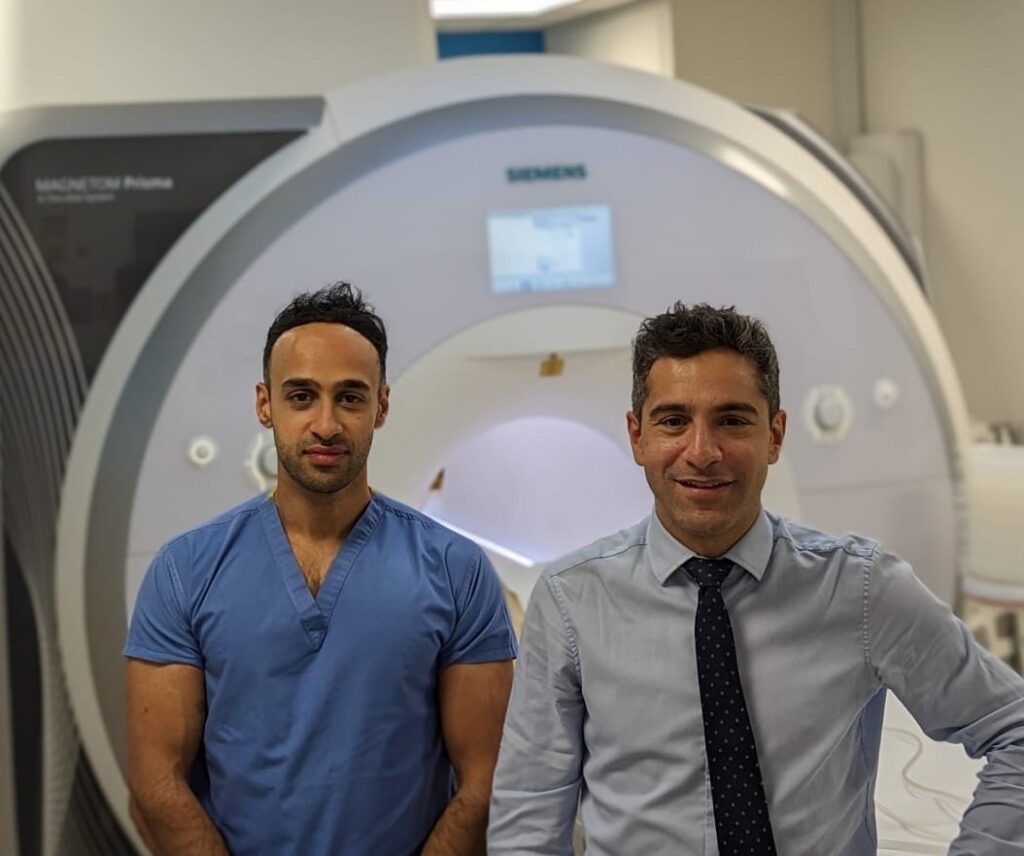
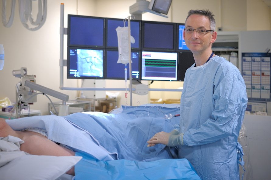
Professor Declan O’Regan
Imperial College London
Amount: £227,898

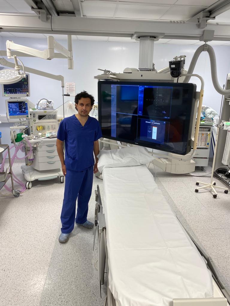
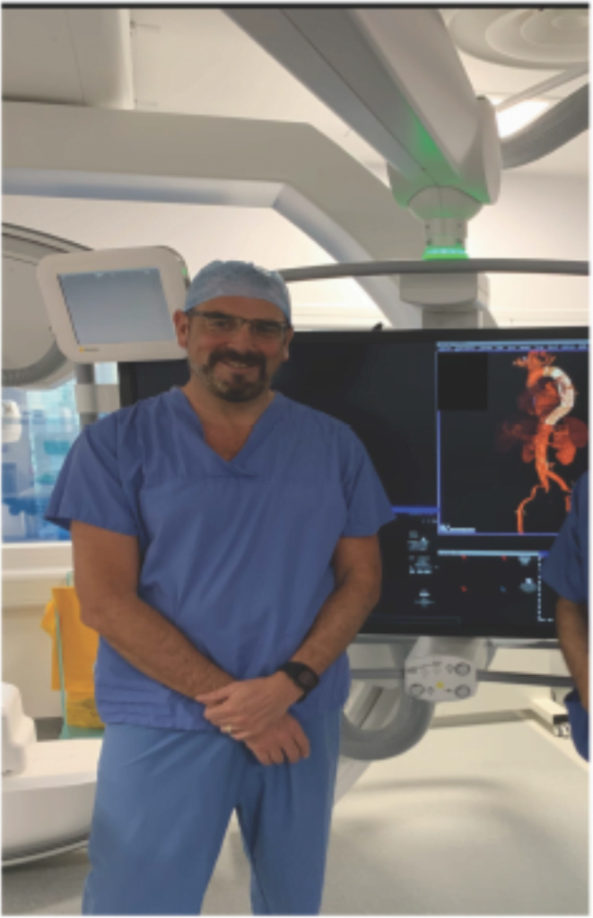
Summary: Thoracic aortic aneurysms can lead to life threatening dissection or rupture of the aorta. Current routine scans and surveillance programmes fail to catch many of these events before they happen, leading to poor outcomes. Professor O’Regan and his team are developing an artificial intelligence system that will generate a 3D model of the aorta and predict the progression of a TAA. If successful, this will help patients and clinicians to make informed decisions about surgery.
The aorta is the main blood vessel in the body, carrying blood away from the heart and around the rest of the body. A thoracic aortic aneurysm (TAA) is a swelling or bulging of the aorta in the chest. Some genetic conditions, such as Marfan syndrome, increase the risk of developing TAA, as do other factors such as high blood pressure or smoking. Although a TAA can remain stable for many years, when they grow beyond a certain point, the individual is at risk for an aortic dissection or rupture. Dissection occurs when the layers of the aorta separate from each other, whereas rupture is when all the layers of the aorta wall tear. Both events are often fatal, and it is therefore of great importance for patients where a TAA is detected that it is monitored closely.
If a TAA is relatively small (between 3 and 5.4cm) then surveillance, involving routine scans, is recommended but if it exceeds this, the patient is usually offered surgery to replace the section of aorta. However, there are two major issues with this. Firstly, the decision to have surgery is one of balance, deciding whether the benefits outweigh the risks. Secondly, 60% of aneurysms have been found to rupture underneath this cut-off point. Given the life-threatening nature of an acute aortic event, improvements in surveillance are required.
Professor O’Regan and his team will use artificial intelligence (AI) technology to analyse CT scans of thoracic aortas and generate 3D images. Not only will this enable more accurate measurement of aneurysms, but it will also allow clinicians to establish which section of the aorta is under the most stress. Additionally, using data from thousands of patients with TAAs, the team hopes that the AI technology will be able to predict which aneurysms are at the greatest risk of dissection or rupture.
This will have major benefits to both patients and healthcare professionals. For patients, it will reduce anxiety surrounding aneurysms and allow them to make informed decisions about surgery if the time comes. For healthcare services, it will enable more accurate and efficient analysis of scans that will greatly benefit the NHS screening and surveillance programmes, ultimately with the view to decrease the number of acute aortic events and save lives.